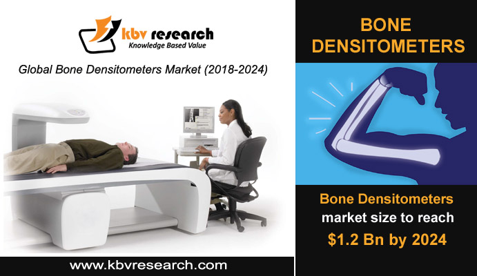
It is anticipated that, by 2040, around 78 million (26%) US adults aged 18 and over will have doctor-diagnosed arthritis. The disease is however found more common among the geriatric population with increasing the risk of arthritis with age. Arthritis is much more prevalent in women than in men. Elderly patients have relatively weaker bones and are more likely to fall as a result of poorer balance, drug side effects and environmental hazards. They are therefore highly prone to bone-related diseases. Physicians rely heavily on bone densitometers to diagnose and treat these medical conditions.
The technology used for bone mass measurement is bone densitometry (also referred to as the dual energy radiation absorption petroleum, DEXA or DXA). It also helps to measure the amount of bone mass in the body of a patient. A narrow, almost invisible, low-dose X-ray beam is used in the bone densitometer. In the scanning process, two energy peaks invade the bones and are used for absorption of soft tissue and for penetration in the bone. Bone densitometer has special software to automatically test and display the results on the computer monitor for the measurement of bone density.
Every few years, several people are tested for bone density. The main reason why the test is being conducted is for severe bone loss (osteoporosis) to be detected and treated and fractures and disability prevention to be prevented. Caring for bone health is a lifetime process.
The human body undergoes a constant process of digging out an old bone and filling it with a new bone, and the whole skeleton takes about 10 years. During this time, all bones are reworked. And what happens in any bone disease is that the patients break down more bones than they form or they form more bones than the required ratio. They break down more bone over time in osteoporosis and it weakens and predisposes it to fracture.
Incorrectly, people often think the topic of bone health and osteoporosis is meant for the elderly. However, it is evident that young people should be more careful about their bones. Possible explanation being, bones peak out around the 30s and then you begin to decline gradually in your bone and muscle. This makes it essential that your bones are checked every now and then thoroughly.
Click Here For Free Insights: https://www.kbvresearch.com/news/bone-densitometers-market-size/
Usually the DEXA scan evaluates bone density or measures it. It also may have applications such as the percentage of magnetic muscle and fat in determining body composition. The DEXA scan is done on two low-energy X-rays, directing doctors towards the bones. The DEXA scanner uses two low energy X-rays.
Utilizing dual energy levels, the physician can separate the images into two components, including soft tissue and bone. If a person has a low bone density or if the condition gets worse, a DEXA scan is much more precise than a typical X-ray because it can detect even the small changes in bone loss.
A Central DXA scan involves lying on a table while your hip, spine and other torso bones are scanned by an X-ray machine. The bones of your forearm, wrist, fingers and heel are tested by a peripheral DXA scan. This scan is usually used to learn if a central DXA is required. The test takes just a couple of minutes.
Since X-rays are used in a bone mineral density test, radiation exposure is a substantial risk. The radiation of the test is, however, very low. Experts agree that the risk posed by this exposure to radiation is much lower than the risk of not detecting osteoporosis before you even have a bone fracture. In addition, excessive radiation exposure always has a small chance of cancer. The advantages of a proper diagnosis, however, far outweigh the risks.
The bone densitometers market has seen a massive adoption recently because the bone densitometry is an accepted standard for bone material density measurement. It is also used to discover osteopenia or osteoporosis that reduces a bone's minerals and density. Speedy advancements in this industry are the result of the growing geriatric population that is extremely vulnerable to osteoarthritis.
In the development of the bone densitometer market, technological advances have played a major role. The implementation of 3D-DXA increased the performance of DXA scans and allowed 3D models of 2D bone images to be constructed in a routine exam. This helps us understand better the state of bone, bone geometry, thickness of the cortical bone and bone minerals. Continuing trends and challenges are expected to boost the Global Bone Densitometer Market collectively with a 4.7% CAGR market growth over the forecast period.